“I am honored and grateful for the time and expertise that Dr. Rajendran has dedicated towards educating us on this very important topic. I feel it is extremely important that everyone should understand the symptoms and the treatments regarding brain metastasis. I hope you find this page to be a resource to empower you with knowledge.” xxooo, Cynthia
The brain is a critical organ that controls many of the parts and functions of the human body. So, naturally when someone who is already fighting breast cancer is diagnosed with brain metastasis, it can be especially devastating to both the patient and their family.
Any patient with breast cancer can potentially present with metastatic disease. While metastasis can develop anywhere in the body, the brain is unfortunately one of the most common sites for those diagnosed with metastatic breast cancer. The most common symptoms of breast cancer-related brain metastases include:
The initial evaluation of brain symptoms includes a complete review of the patient’s medical history and physical exam. Patients also undergo a neurologic examination to evaluate their motor strength, balance, reflexes, sensation, coordination, and cranial nerves.
To better understand the treatment for brain metastases, we will look at three different MRI scans to determine the type of presentation and treatment. The three representative MRI scans will demonstrate the thought patterns for surgical resection versus whole brain radiation therapy versus stereotactic radiation therapy (gamma knife radiosurgery).
Radiation therapy can produce a range of different side effects, so it’s important to discuss them with your neurosurgeon. The most common short-term side effects can include:
Gamma Knife Radiosurgery is a painless computer-guided treatment that delivers highly focused radiation to tumors and lesions in the brain. It is performed by a neurosurgeon while the patient is under local anesthesia.

“My joy is giving the patient a complete understanding of the role of radiation oncology in the treatment of cancer. That knowledge is powerful and pays forward as my patients can teach others.”
Ramji Rajendran MD, PhD
Radiation Oncologist
Sign up for our newsletter.
But it is important to understand what a brain metastasis is and why it’s different from a primary brain tumor. Unlike a primary brain tumor, which forms on the inside of the brain, brain metastasis form on tissues outside of the brain. This classifies them as secondary brain tumors. In some cases, this type of tumor may still respond to breast cancer specific treatments, but due to a special protective mechanism called the blood brain barrier, many breast cancer treatments do not reach the brain. In these types of situations, radiation therapy and surgery remain the most effective treatments for brain metastasis.
A CT scan of the head is performed to ensure that the neurologic symptoms are not being caused by bleeding or trauma and an MRI of the brain (with contrast) is performed to clearly delineate soft tissues within the brain and metastasis.

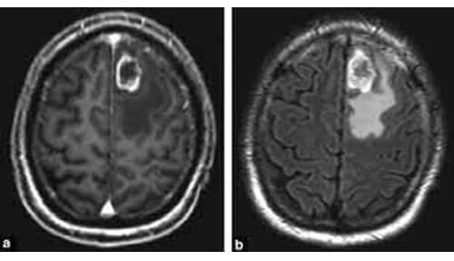
In the images above, we see two MRIs of the brain of the same patient. Panel a demonstrates the tumor volume while panel b demonstrates the swelling caused by the tumor. This patient has a single large metastasis located in a part of the brain that the neurosurgeon determines to be safe, so they are an appropriate candidate for surgery. Surgery allows the tumor to be removed, so it can be further examined to verify that it is, in fact, a metastasis. The neurosurgeon can also examine molecular targets for treatment such as estrogen receptor or Her-2. The patient will undergo radiation therapy for about three weeks after surgery to help prevent the tumor from coming back.

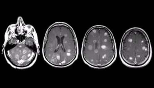
In the images above, we see multiple slices of an MRI of a patient’s brain. These images demonstrate multiple tumors throughout the brain. Since multiple brain metastasis are causing the symptoms, this patient is an appropriate candidate for whole brain radiation therapy. This involves using external beam radiation therapy consisting of ten treatments delivered over two weeks.

In the image above, the MRI shows that the patient has what is classified as “limited” brain metastasis. Limited disease can be determined as either as 1-3 brain metastasis or multiple tiny metastasis. For this patient, Gamma Knife Radiosurgery will be the plan of action to treat the brain metastasis.
The gamma knife allows for very small volumes of tumors to be treated to a very high dose of radiation. This is done by using a machine that emits 192 beams of radiation therapy all focused on one small volume. Gamma Knife Radiosurgery can treat tumors as small as 3 mm in diameter and it can be used to treat 1-3 brain metastasis or low volumes of brain metastasis for select patients that have a good performance status and well-controlled cancer outside the brain.
Here is more information about gamma knife radiosurgery:
Lorem ipsum dolor sit amet, consectetur adipiscing elit. Ut elit tellus, luctus nec ullamcorper mattis, pulvinar dapibus leo.
Learn more