Breast Cancer Surgery
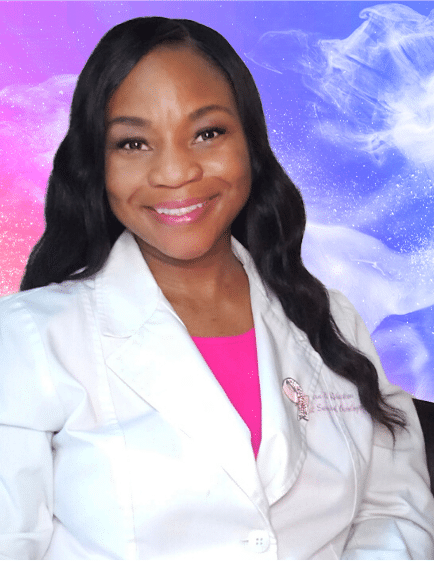
Understanding Breast Cancer Surgery with Dr. Yara Robertson
IN THE KNOW – BEFORE SURGERY
DR. YARA ROBERTSON - BREAST SURGICAL ONCOLOGIST
An informed patient is the best advocate for learning about your breast cancer and treatment option. It can give you back some of your control. Trying to process information and make treatment choices while trying to process your feelings about a breast cancer diagnosis can be overwhelming and disorientating. Take a deep breath, it will be okay.
I am working with Learn Look Locate on this dedicated web page to provide answers to key questions that a person, newly diagnosed with breast cancer might have regarding their upcoming surgery and treatments.
I feel this is a much-needed resource to ease the anxiety of a diagnosis and way to empower someone about their breast cancer treatment.


According to the National Coalition for Cancer Survivorship, “survivorship” starts at the moment of a cancer diagnosis and continues throughout the rest of your life. Some call themselves survivors the moment treatment is complete, some wait until they have been cancer free for a number of years, some call themselves a survivor the day they are diagnosed, and others avoid using the term to describe themselves altogether and prefer to be called “thrivers”. A thriver which focuses more on living well after their diagnosis.
Survivorship is learning to live with, through, and beyond cancer while being able to focus on the ability to cope emotionally and physically with the long-term effects of diagnosis and treatment.
Being a cancer survivor will mean different things for different people and that is ok, because every cancer experience is unique.
A sentinel lymph node is the first lymph node to receive drainage from the breast that could potentially have cancer cells from a primary invasive breast cancer. There are usually 1-3 sentinel lymph nodes.
We check the sentinel lymph node at the time of surgery for invasive breast cancers for both patients undergoing a lumpectomy or mastectomy. We either inject a radioactive tracer, blue dye, or both into the breast or around the area of the tumor.
This helps us to figure out which lymph nodes are the first to receive drainage from the breast and those lymph nodes that could be first to be invaded by breast cancer cells. We typically remove between 1-3 lymph nodes. If the lymph node(s) are positive we may remove the other nodes.
This is called an axillary node dissection. Before the 1990s we removed all the lymph nodes if you were diagnosed with breast cancer.
As you can imagine, a lot of women did not have any cancer in those lymph nodes, but ended up with significant arm swelling also know as lymphedema. With the development of the sentinel lymph node for the breast, we have reduced the morbidity of axillary surgery because we realized that if the sentinel node was negative, it is very highly unlikely the rest of the nodes upstream from the sentinel lymph node would be positive.
The invention of the sentinel lymph node revolutionized the way the surgical oncology community treated breast cancer and has prevented post-surgical complications for so many countless patients.
A breast surgical oncologist is a surgeon who specializes in treating breast cancer and benign diseases (non cancerous) of the breast. Breast surgeons in the United States have completed a surgical residency. Some breast surgeons have completed additional training, called a fellowship, which is dedicated to breast surgery or oncology. If your breast surgeon is not fellowship trained, remember that some surgeons started practicing before breast surgery fellowships were available. Even without a fellowship, specialized breast surgeons have dedicated their professional careers to the disease and perform hundreds of breast cancer surgeries every year. Your breast surgeon should have a comprehensive knowledge of cancer biology, genetics, and be able to provide you with the most modern surgical options to help you achieve the best results possible.
Remember that no one cancer’s journey is the same, ignore the horror stories and focus on your own story! Trying to process information and make treatment choices while trying to process your feelings about a cancer diagnosis can be overwhelming and disorienting. Take a breath. It will be ok.
• When you first meet with the surgeon, bring another pair of ears (family member, trusted friend, etc.) The person you choose should be reliable and knows how to advocate on your behalf.
• Consider bringing a notebook to write information down and if you have a list of questions, consider bringing it with your appointment with you so you can remember what you need to ask. Make sure you understand that you know who is on your team – Surgical Oncologist, Medical Oncologist, Radiation Oncologist, Genetics, Palliative Care, Nutrition, Social Work, etc.
• Make sure you know what type of cancer you have. Don’t be afraid of asking for your pathology report for better understanding. Learn about the treatment options available. After each appointment you should glean more and more information.
• Feel comfortable with your healthcare team. Sometimes this is a lifelong partnership. You should find healthcare providers that will listen to you, that can explain your diagnosis and treatment in a way that you can understand, and who you feel comfortable with.
• Feel free to ask for a 2nd opinion if you want to get one. No one should get offended at your request and you can always return to the original healthcare team once the consult is completed should you choose to.
• Don’t become a Google MD! Limit your online sources and make sure you go to trusted sites about your cancer.
• Don’t be afraid to ask for professional and emotional support if you need to.
Breast cancer begins when a normal breast cell undergoes a mutation process of the genes that control cell growth. Breast cancer cells, like all cells, grow by cellular division. Since each cell division takes 1- 2 months, the process to detect a tumor growing in the body can be between 2 to 5 years. Since tumor cells multiply and divide exponentially—one cell becomes two, two cells become four, and so on—a tumor will increase more rapidly in size the larger it is. According to the Robert W. Franz Cancer Research Center at Providence Portland Medical Center, breast cancer cells need to divide at least 30 times before they are detectable by physical exam. Tumors that are triple negative have greater increases in volume and shorter doubling times than those that are estrogen positive and Her 2 negative. The question of how long a breast cancer has been present in the body when it is diagnosed can be difficult to determine, but it’s likely that many tumors began a minimum of five years before they were detected.
Your doctor has many tests and procedures at their disposal to diagnose breast cancer or to learn if the cancer has spread to other parts of your body, but not all patients need all of these tests and procedures. Your doctor will select the appropriate tests based on your specific circumstances and taking into account new symptoms you may be experiencing. According to the National Cancer Comprehensive Network (NCCN) guidelines, patients with a greater than T2 tumor (>2 cm), or those with node positive disease considering pre-operative systemic therapy (chemotherapy before surgery/endocrine therapy before surgery) the following can be considered:
• CBC (complete blood count)
• CMP (complete metabolic panel) and liver functions and alkaline phosphatase
• CT of the chest/abdomen/pelvis and bone scan
• PET could be considered (optional)
• MRI (optional)
The National Comprehensive Cancer Network (NCCN) discussion of breast MRI recommends the use of MRI for the following clinical indications and applications:
• Staging evaluation to define extent of cancer or presence of multifocal (2 or more tumors in the same quadrant) or multicentric cancer (tumors in other quadrants) in the ipsilateral breast.
• Screening for breast cancer in the opposite breast at time of initial diagnosis.
• Evaluation before and after neoadjuvant therapy to define extent of disease, response to treatment, and potential for breast conserving therapy.
• Detect additional disease in women with mammographically dense breasts.
• To Identify the primary cancer in women who initially present with axillary disease, adenocarcinoma or Paget’s disease of the nipple with a breast primary not identified on mammography, ultrasound, or physical examination.
Breast cancer treatment is individualized and your medical oncologist will look at things such as the size of your tumor, grade of your tumor, the hormone receptors of your tumor, your age, and if there is lymph node involvement to determine if you will need chemotherapy. The goal of chemotherapy is to reduce the risk of cancer coming back. Chemotherapy given before surgery is called neoadjuvant chemotherapy. Your doctor may choose this approach if you have been diagnosed with a locally advanced breast cancer, a triple negative breast cancer, a Her 2 positive breast cancer, or an inflammatory breast cancer. Also, if you have a large breast cancer and want to conserve your breast (lumpectomy), chemotherapy may be given to shrink the tumor before surgery. Shrinking the tumor (or downstaging) allows for less extensive surgery, meaning you may be able to avoid having a mastectomy and/or have a lot of lymph nodes removed in the axilla. Patients with tumors that are small (less than 2 centimeters), low grade, estrogen positive, or Her2-negative may not require systemic chemotherapy and will likely be recommended to have surgery first.
Remember that not all breast cancers are the same and therefore surgical treatment is individualized. Depending on your specific cancer, your surgeon might recommend that you have a particular operation. On the other hand, you might have a choice of operations to consider.
• Lumpectomy/breast conservation therapy is removing the cancer with a surrounding border of normal breast tissue (breast conserving surgery) followed by radiotherapy
• A mastectomy removes the whole breast (mastectomy) and then possibly having a new breast made (breast reconstruction)
•
o If the tumor is larger than 5 cm you will likely need a mastectomy, but in recent years (depending on stage and other factors) patients who under neoadjuvant chemotherapy may be able to shrink the tumor to a size that could be amenable to a lumpectomy. Your doctor will discuss if this is an option for you. Patients with a large amount of DCIS should consider a mastectomy.o If your breast is very small and conserving your breast would potentially leave you disfigured and/or with very little breast tissue, you may be advised to have a mastectomy for a more cosmetically pleasing result.o You may need to consider a mastectomy if you cannot have radiation (had prior breast radiation from a previous lumpectomy, you have a connective tissue disorder like lupus or rheumatoid arthritis, are pregnant, or you don’t want to commit to radiation treatment).o A mastectomy will likely be recommended if you have had multiple surgeries to remove the tumor with lumpectomy but your margins remain positive (the surgeon has not been able to obtain clear margins).At the end of the day, if you feel a mastectomy would give, you’re a better peace of mind than a lumpectomy, have an open and honest discussion with your surgeon about your decision.
Often, I get asked by a patient if they should remove the breast without cancer. Removing the opposite breast without cancer is called a contralateral prophylactic mastectomy (CPM). Research has shown that only 3% to 5% of early breast cancer patients will develop cancer in the opposite breast over the next 10 years after developing breast cancer. Removing the opposite breast offers no survival benefit and does not prevent metastatic disease from developing, except in a very small group of patients. A consensus group from the American Society of Breast Surgeons all agreed that removing the opposite (non-cancerous) breast should be discouraged for the average-risk woman. That being said, your doctor should discuss the benefits and risks of proceeding with removal of the contralateral breast while considering your preference. This is the importance of shared decision making. At the end of the day, you have to make the decision that ultimately you feel comfortable with.
You may require a drain after surgery from your breast cancer. Occasionally a drain is placed after a lumpectomy or limited axillary surgery, but after a mastectomy you will get a drain. Why you ask? Because during surgery the lymphatic vessels are cut removing the breast. These lymphatic vessels normally drain fluid from the breast and after the breast is removed, the fluid can leak into the area that the breast was removed. The body can reabsorb some fluid without any problems, but if this volume of fluid is more than about 25 mL per day the fluid can accumulate and form a seroma.
There are different types of surgical drains that can be used, but mostly like your surgeon will use a Jackson-Pratt. These drains are placed within your surgical field and are attached to flexible tubing that passes through and is stitched to your skin. The tubing is capped with a soft plastic bulb, which catches and holds the fluid, and a stopper outside of your body.
Gradually, the lymphatic vessels will seal off and the drainage should stop. Your surgeon will remove the drain when the fluid is less than 20 ml (but this varies from surgeon to surgeon). This is the reason why it is important to measure the total amount of fluid that is generated through each drain every day so that we can tell whether the drain is ready to be removed. If the drains are removed to soon, the fluid will re-accumulate in the surgical site and it may need to be drained out after surgery.
Yes, no one like drains. They are pesky, annoying, and uncomfortable, but remember: When blood and lymphatic fluid collect in the surgical site, it can cause discomfort, and delay healing if not drained.
A common question that I get asked is how long will my cancer be in the lab?
After surgery for breast cancer, your surgeon sends off the lumpectomy, mastectomy, or lymph nodes for analysis. The specimen(s) go into a container with preservative in most cases. The tissue is then dipped in paraffin wax for protection and cut into very thin slices. These thin slices are placed onto glass slides and stained with dye. The pathologist looks at the slides under the microscope. This is called histology (looks at the cells and tissues). As you can see, this process is not simple, and that is why your official pathology report can take 3-7 days to get back.
By federal law, these slides for histopathology have to be kept for at least 10 years. Cytology is kept for at least 5 years, and paraffin blocks are kept for at least 2 years. Some states have their own laws that require labs to keep pathology specimens longer than required by federal or state laws.


I only wish I had this information and connection with Dr. Yara Robertson before my surgery. I was so nervous, scared, overwhelmed and unsure about what was going to happen. I hope that Dr. Robertson’s soothing words and approach towards this difficult part of the journey gives you some sense of peace. I am so grateful and honored to be working with Dr. Robertson to help fill this much needed void.
XXOO, Cynthia
Dr. Yara Robertson Breast Surgical Oncologist
Educate. Inspire. Connect.
Sign up for our newsletter.

