Search
Search

PATHOLOGY. THE KEY TO YOUR

DIAGNOSIS.

What is a pathology report and why does it matter? Your breast cancer pathology report contains information about your individual case, from the type of tissue being evaluated to your final diagnosis.  Understandably, the terms used in the report can be very complex and difficult to understand, but once I was able to understand what it all meant, it was like a key that unlocked all the information about my breast cancer journey.

I hope this page gives you a patient-friendly approach towards this very important piece of the puzzle of your diagnosis. I am honored and grateful to have Hannah Gilmore MD, Pathologist, provide us all with a roadmap to understanding a breast cancer pathology report. XXOO, Cynthia

Who is a Pathologist?

A pathologist is a medical doctor who has undergone additional training in laboratory techniques used to study disease, most commonly cancers. So, if you are diagnosed with breast cancer, it is the pathologist who made that diagnosis.

Your pathologist is an essential member of your treatment team because he or she will be the one to determine whether your tumor is benign or malignant, as well as how far the tumor has grown within the breast, whether it has spread to lymph nodes or other organs, and other important details.

YOUR TUMOR IS UNIQUE . JUST LIKE YOU.

So, my tumor was very unique, just like me. Everyone’s tumor is distinctly different, so a pathology report is a way to get to know the personality of your tumor on every level so your medical team can determine the best treatment plan for you.“

When you are diagnosed with a lump in your breast, your pathologist takes tissue samples of your tumor, also called a biopsy. This is so they can determine exactly which type of breast cancer you have. The answers to this test are then provided on a pathology report, so the pathologist and the rest of your treatment team will understand your unique case and how best to treat it.

The first three sections on the report provide basic information regarding the Patient, the Physician’s Details of the physician performing the biopsy, and the Specimen Case Number (including the date of the biopsy and the date when the tissue was processed by the lab).

The most important sections are those that follow, starting with Body Site and Procedure. This section details the exact location from where the tissue sample is taken and the procedure that was performed, usually a core needle biopsy, but it could be a more advanced procedure, such as a lumpectomy or mastectomy.

The report also features a section called Gross Description. This section is where the pathologist describes the physical characteristics of the tissue that was removed before they look at it under a microscope. Recorded details can include the size dimensions of the tissue; the colors of ink used to orient the margins of the specimen; the size and location of any visible masses; the distance of the mass to the margins; and the number of sections submitted from the tissue. The amount of information in this section is determined by the type of procedure that was performed. Typically, a lumpectomy or mastectomy will provide substantially more information than a core needle biopsy.

The key to your treatment.

Your biopsy and resulting pathology report are essential because they inform the pathologist and the rest of your treatment team of your diagnosis, including your unique tumor type, tumor grade, tumor stage, and tumor expression of predictive and prognostic biomarkers such as ER (the protein responsible for binding to and detecting estrogen), PR (the protein responsible for binding to and detecting progesterone), and HER-2 (the protein found on the surface of all cells that signal the cells to grow). With this information, they can more accurately determine the best course of treatment for your case.

Unlocking your diagnosis.

The Diagnosis section on your pathology report is the most important part because this is where you will find out if your tumor is benign (not cancerous) or malignant (cancerous). If your tumor is malignant, then the report will also state what type of cancer you have, the most common being carcinoma.

Carcinomas are cancers that arise from the epithelial cells of the breast and can be either ductal carcinoma in situ (DCIS), invasive ductal carcinoma (IDC),  lobular carcinoma in situ (LCIS), or invasive lobular carcinoma (ILC).

Ductal carcinoma in situ is the most common type of noninvasive breast cancer and it starts in the milk duct. It is considered noninvasive because the cancer hasn’t spread from the ducts to any other part of the body.

Invasive ductal carcinoma starts in the milk duct but then spreads to other tissues, including the lymph nodes. If not treated, this type of cancer could metastasize to other parts of the body, such as the bones or brain. Approximately 80% of all breast carcinomas are invasive ductal carcinoma, making it the most common type of breast cancer diagnosed.

Lobular carcinoma in situ is not technically considered a cancer, but it does place a woman at higher risk of developing invasive breast cancer. LCIS is diagnosed when cancer-like cells develop in the breast’s lobules (the milk-producing glands). These cells typically remain in place and do not spread to other tissues.

Invasive lobular carcinoma accounts for 10% of all breast cancer diagnoses. It starts in the lobules and the cancer cells then spread to other parts of the body. Approximately 20% of women diagnosed with ILCS have cancer in both breasts.

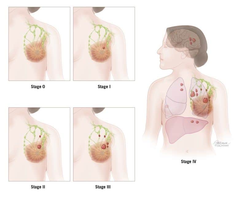
In addition to your breast cancer diagnosis, this section will also tell you the pathologic Stage and Grade of your tumor. The stage of your cancer indicates how advanced it is, ranging from Stage 0 (pre-invasive disease) to Stage IV (metastatic disease). The higher the stage, the more advanced the cancer is.

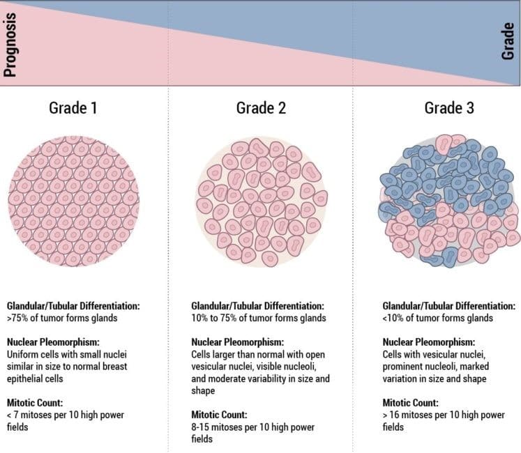
The grade represents the tumor’s “aggressive potential.” Cancers that are rated with lower grades tend to be less aggressive than those with higher grades. Determining the stage and grade is thus very important, because these factors are used to predict your prognosis and will help direct your treatment options.
Also included in the Diagnosis section are details pertaining to whether your cancer is sensitive to your body’s naturally occurring hormones, estrogen and progesterone. This is indicated by Hormone Status. Knowing this information is important because it gives your doctor a better idea of how best to treat the cancer or prevent it from recurring. Hormone status of breast cancers includes:
  • Estrogen Receptor (ER) Positive – If your pathology report states your hormone status as ER Positive, then this means that your cancer is sensitive to estrogen. As such, treatment should include anti-estrogen hormone (endocrine) therapy to block the growth of cancer cells.
  • Progesterone Receptor (PR) Positive – If your pathology report states your hormone status as PR Positive, then this means that your cancer is sensitive to progesterone. Treatment with endocrine therapy will also be applied here to help block the growth of cancer cells.
  • Hormone Receptor (HR) Negative – If your hormone status is HR Negative, then this means that your cancer does not have hormone receptors. Thus, endocrine treatments won’t be effective against this type of cancer and other treatment options will need to be utilized.
  • HER2 Positive – If your pathology report states your cancer as HER2 Positive, then this means your cancer tests positive for a protein called human epidermal growth factor receptor 2. This happens in about 1 of every 5 breast cancer cases. While HER2 Positive cancers are considered more aggressive, their treatments are highly effective, so the prognosis of this diagnosis tends to be good when diagnosed early.
Breast cancer that is identified as ER Negative, PR Negative, and HER2 Negative is what is known as Triple Negative breast cancer. This is considered among the most aggressive types of breast cancer and one of the most challenging to treat. Approximately 10% to 15% of breast cancer diagnoses are Triple Negative. Your doctor will likely order further testing on your cancer if it estrogen receptor positive/HER negative. This is usually done by outside pathology labs. Lastly, the pathology report concludes with the name of your pathologist and their contact information.

PATHOLOGY - THE KEY
TO YOUR DIAGNOSIS

A breast cancer diagnosis can change your life is so many ways, but as with any obstacle you face, the more you know about it, the better you can prepare for what’s ahead. Getting a biopsy and reading your pathology report can be scary but educating yourself will help you better understand the pathology of your tumor. It is also important to know that you are not alone. Connecting with others who are on this same journey can also help you through this challenging time.

Look Learn Locate is designed to help you learn more about your cancer straight from medical professionals and connect with a wider community of cancer patients and survivors. Join our community today to Educate, Inspire, and Connect.

Dr. Hannah L. Gilmore MD

Dr. Gilmore is a board-certified anatomic pathologist specializing in breast pathology at University Hospitals Cleveland Medical Center and Associate Professor of Pathology at Case Western Reserve University in Cleveland, Ohio. 

Dr. Hannah L. Gilmore is a pathologist in Cleveland, Ohio and is affiliated with multiple hospitals in the area, including Louis Stokes Cleveland Veterans Affairs Medical Center and University Hospitals Cleveland Medical Center. She received her medical degree from Case Western Reserve University School of Medicine and has been in practice between 11-20 years. Dr. Hannah L. Gilmore is a pathologist in Cleveland, Ohio and is affiliated with multiple hospitals in the area, including Louis Stokes Cleveland Veterans Affairs Medical Center and University Hospitals Cleveland Medical Center. She received her medical degree from Case Western Reserve University School of Medicine and has been in practice between 11-20 years.

(Image source: https://pathology.jhu.edu/build/assets/breast/_lgImg/breast-cancer-stages-illustration.jpg)

(Image source: https://pathology.jhu.edu/build/assets/breast/_lgImg/breast-cancer-grades-illustration.jpg)

(Image source: https://pathology.jhu.edu/build/assets/breast/_lgImg/breast-cancer-grades-illustration.jpg)

(Image source: https://pathology.jhu.edu/build/assets/breast/_lgImg/breast-cancer-stages-illustration.jpg)